Comment on the interconnected interstitial system by Robert Schleip
This is the second article concerning fascia from the group around Neil Theise (New York) appearing in a high-ranking journal of the Nature publishing group. In the first paper, published in 2018, they reported their discovery of a loose connective tissue network containing a fluid-filled reticular architecture in the submucosa of the gastrointestinal tract and several other places (Benias et al. 2018, Sci. Rep 8: 4947).
This first publication elicited both enthusiastic as well as critical responses internationally. The critical responses mainly were related to the relative lack of recognition within this article that several other authors had previously described this ‚interstitial network‘, such as Jean-Claude Guimberteau or Andrew Taylor Still, although their descriptions had not become part of standard textbook anatomy.
This first paper could not answer one question: whether these interstitial spaces are all connected, with an easy fluid exchange capacity between them, or whether some of these spaces are kept apart from each other by separating dense fascial membranes between them. In particular, several fascia-inspired investigators like myself had been wondering to what degree the loose areolar connective tissue under the skin in us humans is kept separate – or openly connected – with the loose connective tissue surrounding our viscera. I had suspected that an easy and unobstructed liquid connection might exist between these „two oceans“ via the fascial connections between the anterior neck and the mediastinum. However, vague speculation is something very different than scientific evidence, derived by robust experimental measurements!
Therefore, reading this brilliant article triggered some great delight in me, as it shows that the different oceans are indeed intimately connected. The authors used several dying methods, such as tattoo pigments, colloidal silver, and a binding protein for hyaluronan, to trace their distribution across different spaces. The results convincingly proved “that interstitial spaces are continuous between tissue compartments and fascial planes in the colon, skin, and liver, as well as within the fibrous tissues around blood vessels and nerves, which may pass through multiple organs“.
Based on this, they suggest “that there is continuity of fibrous tissue interstitial spaces within and between organs and that these spaces are also potentially continuous between more distant parts of the body, at least along the vasculature and nerves. We speculate that these spaces serve as pathways for molecular signalling and cell trafficking in a way that is both in series and in parallel with the established pathways of the cardiovascular and lymphatic systems“.
The authors continue to suggest that this novel clarification may go along with a different spreading concept of cancer cells throughout the body along with this interstitial space (and in parallel to their well-known migration along blood and lymph vessels). It may also be with a more frequent migratory activity of different microbes from our microbiome throughout this body-wide interstitial system. This continuum may shed some new light on the so-called “gut brain axis”. The authors suggest that perineurial fascial structures around the carotid arteries may play a significant role. Given that the volume of interstitial fluid in our body is more than three times the combined fluid volume of the lymphatic and cardiovascular systems, this new finding will most likely inspire other research groups worldwide in the next few years to continue to explore what degree – and along which interstitial channels – such spreading of cells and micro-organisms is actually happening, and how our immune system regulates the constant interaction with them. If you thought you knew what fascia is or that you could understand most of its features by dissecting dead bodies, this article is a beautiful wake-up call. Most possibly, fascia is more alive than we had ever thought!
People with low back pain walk differently
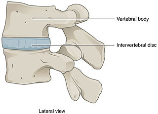
Running exercise strengthens the intervertebral disc