Differences in Knee and Hip Adduction in Runners with Iliotibial Band Syndrome
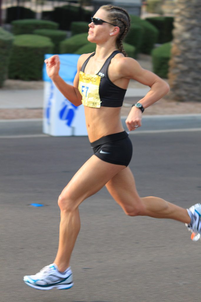
 Iliotibial Band Syndrome (ITBS) has been associated with altered hip and knee kinematics in runners. A previous research identified increased strain rate in female runners with ITBS, and another study demonstrated increased knee adduction (genu varum) in male runners with ITBS. There has been a suggestion that neuromuscular factors at the hip may play an important role in iliotibial band syndrome syndrome.
A study compared hip surface electromyography, and frontal plane hip and knee kinematics, in runners with and without iliotibial band syndrome.
The study was an observational cross-sectional study. Thirty subjects were recruited consisting of 15 injured runners with iliotibial band syndrome and 15 gender, age and body mass index matched controls. In each group, eight were male runners and seven were female runners. Inclusion criteria for the injured group were pain within two months related to iliotibial band syndrome and a positive Noble compression test.
Three-dimensional motion capture was performed using ten high speed cameras synchronized with wireless surface electromyography during a 30-minute run. The first data point was at three minutes using a constant speed of 2.74 meters per second. A second data point was at 30 minutes using a self-selected pace by the participant to allow for a challenging run until completion at 30 minutes.
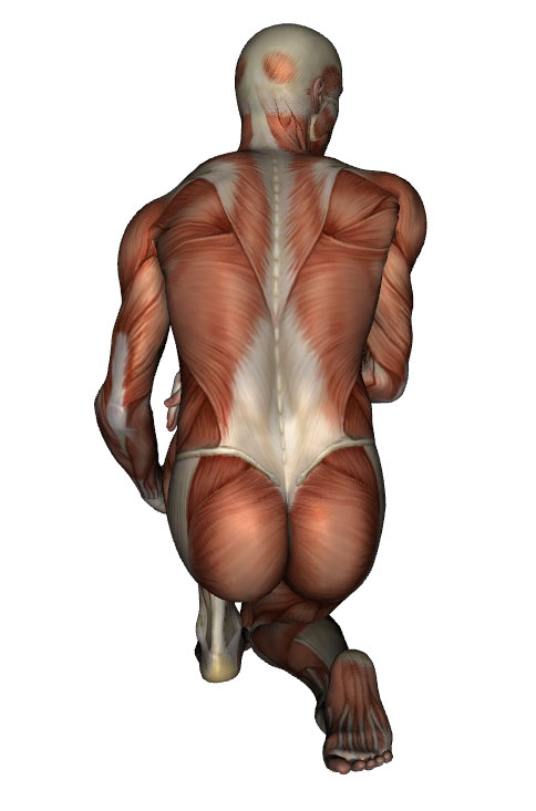
Motion capture was reported as peak kinematic values from heel strike to peak knee flexion for hip adduction and knee adduction (genu varum – bow leg). Surface electromyography was reported as a percentage of maximal voluntary contraction for the gluteus maximus, gluteus medius, and tensor fascia latae muscles.
The data showed that injured runners showed increased knee adduction compared to control runners at 30 minutes (control = -1.48°, injured = 3.74°).
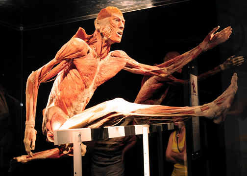
Tensor fasciae latae muscle activation in injured runners was increased compared to control runners at three minutes (control = 7% MVIC, Injured = 11% MVIC).
The authors suggested that lateral knee pain in runners localized to the distal iliotibial band is associated with increased knee adduction at 30 minutes. Increased Tensor Fasciae Latae (TFL) muscle activation at three minutes is noted but more investigation is needed to better understand the clinical meaning. These findings are consistent with but not conclusive evidence supporting the theory that neuromuscular factors of the hip muscles may contribute to increased knee adduction in runners with iliotibial band syndrome.
Nevertheless, the authors advise caution using these findings to support treatments intended to modify TFL activation given the small differences of four percent in muscle activation. Increased knee adduction in runners at 30 minutes was over five degrees and beyond the minimal detectable difference. Additional research is needed to confirm if the degree of knee adduction changes earlier versus later in a run, and whether or not fatigue is a clinically relevant factor.
Outside of the published data, the main author of the research, Bob Baker said that the fascial system likely experiences a strain and abnormal strain rate. This affects the pain receptors (i.e., tender) and mechanoreceptors (i.e., muscular activity abnormal in region). For the soft tissue person, this adds the perspective of normalizing these receptors so the pain receptors calm down and mechanical receptors start to fire. I believe this happens during the soft tissue work with various approaches at reducing swelling, pushing out neuromodulators and manipulating deep fascia. Therapists could then try to impact the movement system with movement cues like better diaphragmatic breathing with walking and sport.

