Exercise attenuates fibrosis to the multifidus muscle associated with intervertebral disc degeneration
 The International Society for the Study of the Lumbar Spine recently awarded the ISSLS Prize in Basic Science 2019 to a paper by G. James, Paul Hodges and colleagues from the University of Queensland. The study, published in the European Spine Journal, found that Intervertebral disc degeneration is associated with fibrosis in the multifidus muscle and exercise attenuates that fibrosis
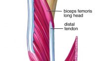
Chronic low back pain is a major disability in society. Low back pain is usually accompanied by structural remodelling and inflammation of muscles around the spine, in particular, the multifidus muscle. Studies have found an increased cross-sectional area of connective tissue and expression of collagen in the multifidus during early chronic LBP. These changes are characteristic of tissue fibrosis. However, the extent and mechanisms underlying the increased fibrotic activity in the multifidus are unknown.
Physical activity can modify connective tissue in skeletal muscles. Short-term exercise stimulates both collagen synthesis and degradation which assist in its remodelling. Long-term exercise prevents aging-dependent fibrosis. A past study found that physical activity reduces local inflammation that precedes multifidus fibrosis during intervertebral disc degeneration (IDD).
Thus this new study aimed to evaluate the development of fibrosis and its underlying genetic network during IDD and the impact of physical activity.
The study used mice that were either sedentary or housed with a running wheel, to allow voluntary physical activity. At 12 months of age, IDD was assessed with MRI, and multifidus muscle samples were harvested from L2 to L6.
The study found:
- Fibrosis (e., increased thickness of the connective tissue between the multifidus and longissimus muscles) in muscle that crossed a degenerated disc.
- Fibrotic gene network (CTGF, SP, TIMP1, and TIMP2) was dysregulated in multifidus crossing a degenerated disc and correlated with changes in Extra Cellular Matrix (ECM) gene expression
- Physical activity attenuated the IDD-dependent increased connective tissue thickness and reduced the expression of collagen-I, fibronectin, CTGF, substance P, MMP2 and TIMP2 in mice.
The authors concluded that the fibrotic networks that promote fibrosis in the multifidus muscle during chronic IDD. Furthermore, physical activity is shown to reduce fibrosis and regulate the fibrotic gene network.
Comments by Til Luchau:
Some degree of connective tissue fibrosity is a normal result of inflammatory reactions to injury—scar tissue is one example. But when inflammatory reactions are extreme or prolonged, excessive fibrosity and pathological, painful scarring play into a vicious cycle of less movement; more sensitivity and pain; and more inflammatory response.
Our field is in the midst of a debate between differing points of view about tissue and pain. Is it tissue density that causes pain (the conventional working assumption behind many manual therapies)? Or is it that tissue fibrosity is an unrelated by-product of pain-induced stasis?
However, there is widespread agreement, across many points of view and therapeutic disciplines, that movement often helps with injury recovery: especially active, moderate and regular movement. This recent study confirms that (at least in SPARC-null mice, which are genetically prone to disc injuries) there are indeed biological, tissue-based differences, and better healing, that result from voluntary active movement after an injury. But far from confirming whether fibrosity is the injury-chicken or the pain-egg, it reveals the intrinsic interconnection between our tissues, our activities, and healing from an injury.
Massage Therapy Decreases Cancer-Related Fatigue