Fascia and the Mind-Body Connection by David Lesondak
That there is a distinct relationship between thought, emotion, and the body should be apparent to anyone involved in the realm of fascia-oriented bodywork. For some, the idea that we are actually working with fascia is controversial but for the purposes of the next 1,420 words let’s accept that we are doing just that. So what is it about fascia therapies that have the potential to induce such strong emotional expressions? For as long as I can remember I have been fascinated by this relationship. And there’s a lot of interesting things out there that begin to explain the pathways of just how such a thing is possible.
One of the most startling is the existence of the myodural bridge1,2. The myodural bridge is a literal connection – a fascial, fibrous, physical connection between the dura mater the sub occipital group. Further investigations reveal the existence of proprioceptive nerve endings in the myodural bridge and suggests both the regulation of dural tension and cerebrospinal fluid flow3.
Someone, somewhere, said “What you don’t express, you repress.” Repression takes energy. Repression creates tension. Think back to the last time you really wanted to say something but were afraid, or really wanted give someone a piece of your mind but didn’t. Was it easy to modulate your emotions in those circumstances? Did it create a fair amount of tension in your body? If you’re being honest, I would wager it did.
So lets go into the extreme and think about the physiology of the startle response, particularly the contraction of the neck muscles. And while it is possible, albeit difficult, to regulate the startle response, it is considered innate. So innate that it is referred to as the Moro Reflex in newborn babies. Fold in anxiety-induced startle response, PTSD, and a dash of fear. We’ve all seen people like that. Is it any wonder that when that tension releases, however momentary or lasting, that there can be a concurrent expression of emotion?
I’m not suggesting a simple cause and effect here, release the sub occipitals and release the trauma. If only if were that easy. We’d all just get a Stillpoint inducer, then Netflix and chill as needed. Life and the body is a little more complicated than that. Let’s go back to the anatomy.
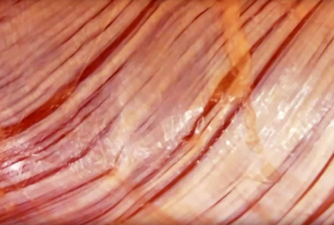
The dura mater is part and parcel of the fascial system, often specifically referred to as the meningeal fascia. The meningeal fascia covers and interpenetrates the nerves. It includes the dura, arachnoid, and the pia mater. The pia is the finest expression of the neural aspect of the fascial system, following all the twists, turns, and contours of the brain all the way down to the ependyma, where the cerebrospinal fluid is produced.
The individual collagen fiber direction in the pia are unidirectional. Some think it capable of communicating tensile forces throughout the meningeal network4. And there’s another intriguing study indicating a relationship between collagen laxity and anxiety5. This makes me wonder if there might be a natural level of pre-tension in the brain, just as there is in the body.
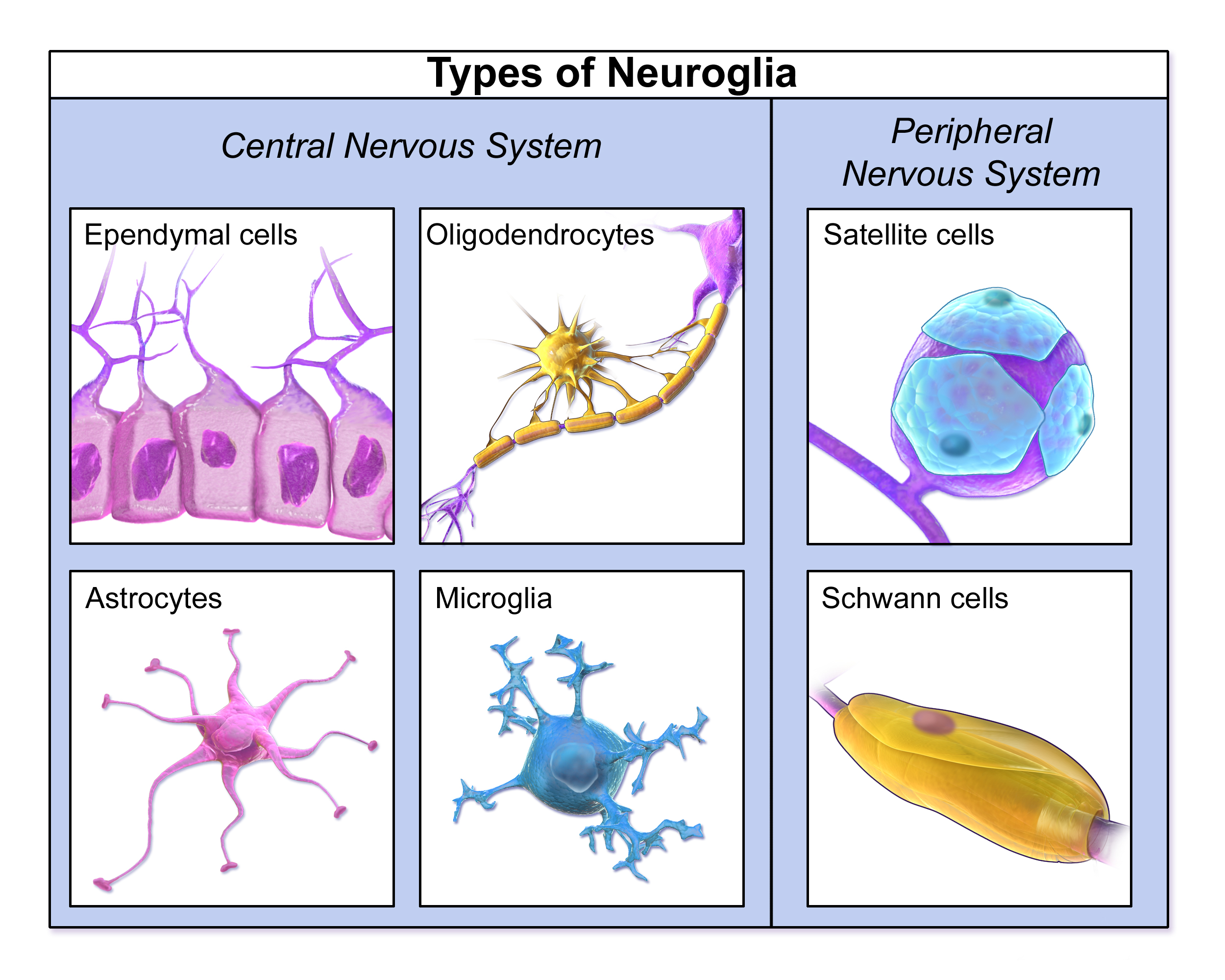
Speculation aside, a vital player in this game is a class of cell known as the glia. And the story of the glia has curious parallels to the story of fascia and the fascial network6. And in terms of tension, glial cells also possess integrins, the cell receptors that respond to pressure and vibration.
A neurological outlier, glia cells outnumber the neurons nine to one in humans (animals have a lower ratio). They were ignored in the early days of neuroscience in favor of the neuron which were less abundant but far larger. In an interesting example of confirmation bias, in this case that bigger is better, they were ignored in favor of the neurons. Glia were thought to be inert, mere structural support and scaffolding for the neurons. Sound familiar?
That changed in the early 1990s when it discovered that not only are the glia not inert, but that they communicate to each other and to the neurons7. Put another way, the cells previously thought to be the stuffing between the neurons are talking to each other. Since then there has been an explosion in glia research, with discoveries that they can regulate synaptic activity8, may be essential to the formation of muscle memory9, and play a role in chronic pain10. In fact, prolonged exposure to opioids cause glia to release pro inflammatory agents11. Glia are also attributed, by the their top researchers, to imbuing the body with the ability to move and perform physical tasks with grace and ease. This too, has a familiar ring.
Most of the literature divides the glia into 4 types (though some of the literature will refer to only 3) the one I want to focus our attention on is the Schwann cell, the only glia cell to arise from the mesoderm, the same embryological layer as our connective tissue.
Schwann cells are found in the peripheral nervous system, and while there are at least 3 distinct types, the ones that hold my interest the most are the perisynaptic Schwann cells (PSCs). PSCs live in the neuromuscular junctions, where the motor commands of the central nervous system are performed. PSCs are considered indispensable to short-term plasticity in the neuromuscular junction12. It is here where the Schwann cells interact with fascial mechanoreceptors, rather like they do at the myodural bridge. PSCs are vital to the healthy formation of both Golgi and Pacini receptors, and where PSCs are absent, these mechanoreceptors cannot regenerate after injury. They have an interdependent relationship13.
Just as we are beginning to map the fascial pathways of force transmission in the muscles and bones (the musculobonular system?) so too I believe we are discovering the pathways that more intimately link our mind, body, and emotions via the pathway just described.
But corollary is not causation, nor is association attestation. It is my fervent wish that some researcher(s) somewhere will begin to put some of these pieces together. It has been my direct experience that glia scientists are ignorant of fascia science, and why should it be otherwise? Our own silos are so deep and rich that it is easy to stay happily inside them.
However, I believe the deeper one digs into the mechanisms between the mind and the body, one is left with the realization that this unitary relationship is so. That it is ineluctably interconnected. And while it is useful, even necessary, to separate and isolate things for study, that we are irrefutable one. And that the fascia seems to be the interface of that oneness.
Fascia What is and Why it Matters by David Lesondak available at www.terrarosa.com.au
David Lesondak, BCSI, ATSI, FST, FFT, VMT, is the author of the international bestseller, Fascia What it is and Why it Matters (Handspring 2017). He is a member of the Allied Health professional staff in the Department of Family and Community Medicine at the University of Pittsburgh Medical Center. A Fascia Specialist in UPMC’s Center for Integrative Medicine, David lectures and teaches hands-on course worldwide. For more information www.fasciamatters.health
References:
1) Von Lanz T. Uber die Ruchensmarkshaute. I. (1929) Die konstruktive Form der harten Haut des menschlichen Ruckenmarkes und ihrer Bander. (The structural form of the hard skin of the human spinal cord and its bands). Arch Entwickl Mech Org 118:252–307
2) Khan j l, Sick h and Kortike J G (1992) Les espaces intervertébraux postérieurs de la jointure craniorachidienne. Acta Anat (Basel). 144 (1) 65-70
3) Scali F, Pontell M E, Enix D E and Marshall E (2013) Histological Analysis of the rectus capitis major’s myodural bridge. The Spine Journal. May; 13 (5) 558-563
4) Nam MH, Baek M, 2014, Discovery of a novel fibrous tissue in the spinal pia mater by polarized light microscopy. Connect Tissue Res. 2014 Apr;55(2): pp. 147–155
5) Bulbena A, Gago J, Sperry l, and Berge D (2006) The relationship between frequency and intensity of of fears and a collagen condition. Depress Anxiety, July;23 (7) 412-417.
6) Lesondak, D. (2017) Fascia and the Brain, Fascia: What it is and Why it Matters, Handspring Publishing, pp. 87-104
7) Nedergaard, M, (1994) Direct signaling from astrocytes to neurons in cultures of mammalian brain cells. Science, Mar 25;263(5154):1768-71
8) Eroglu C and Barnes, B A (2010) Regulation of synaptic activity by glia. Nature. November; 468, 223–231
9) Hassanpoor H, Fallah A and Raza M (2012) New role for astroglia in learning: Formation of muscle memory. Medical Hypothesis. December; 79(6) 770–773
10) Fields R D (2009) New culprits in chronic pain, Scientific American. November; 50–57.
11) Johnston I N, Milligan E D, Wieseler-Frank J et al. (2004) A role for proinflammatory cytokines and fractalkine in analgesia, tolerance and subsequent pain facilitation induced by chronic intrathecal morphine. J Neurosci. August; 24 (33) 7353–7365
12) Colomar, A, Robitaile, R., Glia Modua Costandi, Mo, (2012), Snapshots explore Einstein’s unusual brain, Nature News (online) Nov. 16, 2012
13) Kopp D M, Trachtenberg J T and Thompson W J. (1997) Glial growth factor rescues Schwann cells of mechanoreceptors from denervation-induced apoptosis. J Neurosci. September; 17 (17) 6697–6706
Fascia-focused movement criteria