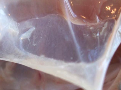
Fascia can actively contract and thereby influence musculoskeletal dynamics
Fascia is a biological fabric that enmeshes all structures in our body. While there is a research interest in the role of fascia as a force transmitter in muscular dynamics, fascia is often regarded as a passive contributor to biomechanical behaviour.
There have been several studies that indicated the active role of fascia which has an inherent ability to contract actively. These indications include the reported phenomenon of “ligament contraction” of human lumbar fascia in response to repeated isometric strain application. There is also evidence of fascial tissues can shorten over several days in certain pathologies, such as Palmar fibromatosis, hypertrophic scars, and similar fascial fibrotic conditions. This tissue shortening is mostly due to the presence of myofibroblasts (a type of cell responsible for wound healing and tissue repair). The resulting tissue contracture is due to an incremental combination of cellular contraction, collagen cross-linking and matrix remodelling.
Robert Schleip and colleagues from Ulm University has been interested in finding out whether normal fascia may possess the capacity for cellular contraction which, in turn, could play an active role in musculoskeletal mechanics.
In a new study published in Frontiers of Physiology, they studied human and rat fascial specimens from different body sites for the presence of myofibroblasts using immunohistochemical staining for α-smooth muscle actin (n= 31 donors, n=20 animals). Also, mechanographic force registrations were performed on isolated rat fascial tissues which were exposed to pharmacological stimulants to measure contracting force.
The study found that
- the density of myofibroblasts is larger in the human lumbar fascia in comparison to fasciae from the two other regions examined in this study: fascia lata and plantar fascia.
- Fascial tissues contract when exposed to different pharmacological substances: fetal bovine serum, the thromboxane A2 analog U46619, TGF-β1, and mepyramine.
- Botulinum toxin type C3–used as a Rho kinase inhibitor– provoked relaxation.
- In contrast, fascial tissues were insensitive to angiotensin II and caffeine.
- There is a positive correlation between myofibroblast density and contractile response.
The calculation of potential contractile forces of fascia predicts a force range that seems insufficient for exerting a direct short-term effect (i.e., occurring within minutes to hours) on mechanical joint stability of the human spine. The short-term contractile forces of fascial tissues are at least two orders of magnitude below that of muscle tissue and may have no significant effect on spinal stability or other important aspects of human biomechanics.
Nevertheless, the predicted fascial contraction forces in the human lumbar region are above the much lower threshold for influencing mechanosensation. They are strong enough to alter motoneuronal coordination in the lumbar region. In addition, the authors suggest that a local and/or temporal increase in fascial contractility might contribute to long-term tissue contracture, which includes matrix remodeling.
Based on the known signaling influence of the sympathetic nervous system on TGF-β1 expression, they suggest that their findings tend to support the hypothesis of a close connection between fascial stiffness and chronic sympathetic activation. In the light of the large contribution of psychosocial factors in low back pain, they suggest further studies to explore possible interactions between emotional stress, fascial stiffness, and low back pain.
The authors concluded that the tension of myofascial tissue is actively regulated by myofibroblasts with the potential to impact active musculoskeletal dynamics.

