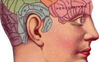
Fasciae around the median nerve could influence carpal tunnel syndrome
Carpal tunnel syndrome the most frequent debilitating condition of the hand, affecting 1 to 3% of the population. It is commonly described as the entrapment of the median nerve at the carpal tunnel. There is also a hypothesis that it is due to compression in the nerves serving the epineurium (outermost layer of connective tissue surrounding a peripheral nerve) and the median nerve, the double crush syndrome.
Carla Stecco and colleagues from Padova University in Italy in a study, investigated the anatomical connections between the median nerve and surrounding structures, which could contribute to compression of the nerve and may play a role in the carpal tunnel syndrome.
The researchers took four samples of the median nerve and surrounding tissues from nine non‐embalmed upper limbs for analysis using microscope. Additionally, ultrasound images were taken from 21 healthy subjects and 16 carpal tunnel syndrome patients to evaluate median nerve transversal displacement during finger motion at carpal tunnel and forearm levels.
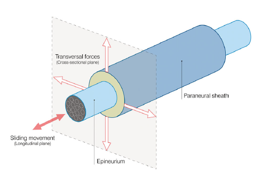
The researchers found an anatomical continuity between epimysium (outermost muscle connective tissue) and the paraneural sheet (outer nerve layer)/ There is a reduction of paraneural fat tissue from proximal to distal in all cadaver samples. Paraneural compartment is a highly organized fibro-adipose structure with a protective role against compression forces.
In the ultrasound study of human, median nerve displacements at both carpal tunnel and forearm levels were significantly reduced in carpal tunnel syndrome subjects.
They found that the median nerve is not an isolated structure but is entirely connected to myofascial structures. The nerve cannot be considered as independent, but embedded in a synergic structure, with the surrounding tissues which participate in its movement.
The researchers propose a 3D model of median nerve and paraneural compartment.
In normal conditions, transversal forces put tension on the paraneural sheath, keeping functional space of nerve open (nerve area and paraneural compartment). Sliding motion occurs due to the gliding surfaces of the connective tissue. When the connective tissue is impaired, this sheath is disrupted due to altered myofascial transmission. This condition may lead to a reduction in functional space, affecting the nerve and increasing shear stress between connective layers.
Spanish Squat Exercise

Massage therapy and exercise are more effective than pain education for chronic neck pain