Intermuscular force transmission along the myofascial meridians
Frieder Krause and colleagues from Department of Sports Medicine, Goethe University in Germany published a review in Journal of Anatomy to provide an evidence on tensile transmission along myofascial chains based on anatomical dissection studies and in vivo experiments.
As evidence for the existence of myofascial chains is growing, and the capability offorce transmission via myofascial chains has been hypothesized. However, there is still a lack of evidence concerning the functional significance and capability forforce transfer.
A systematic literature research was conducted using scientific publicationn databases. The study included myofascial meridians of the superficial backline (SBL), the back functional line (BFL) and the front functional line (FFL). Peer-reviewed human dissection studies as well as in vivo experiments reporting intermuscular tension transfer between the constituents of a myofascial chain were included. To assess methodic quality, two independent investigators rated studies by means of validated assessment tools. The literature research identified 1022 articles. Nine studies (moderate to excellent methodological quality) were included.
And the finding is…
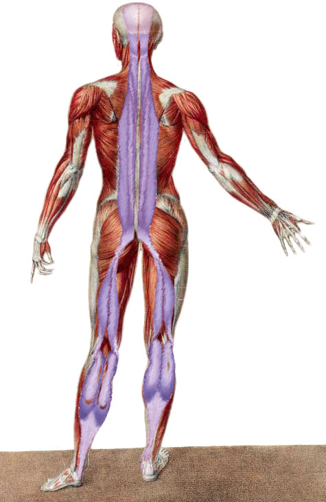
Superficial Back Line
Two studies reporting force transfer between the plantar fascia and the Achilles tendon were found (moderate evidence). One trial identified force transfer between pelvic motion/hamstring movement and gastrocnemius muscle (moderate evidence), and three studies demonstrated force transfer between the hamstrings and the sacrotuberous ligament or the TLF, respectively (moderate evidence).
Back Functional Line
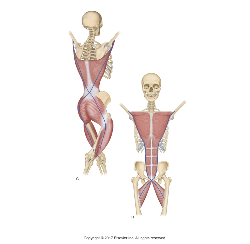
BACK AND FRONT FUNCTIONAL LINE MYOFASCIAL CHAINS. PERMISSION JOSEPH E. MUSCOLINO. KINESIOLOGY – THE SKELETAL SYSTEM AND MUSCLE FUNCTION, 3RD ED. (ELSEVIER, 2017).
The Back Functional Line begins with the latissimus dorsi, connects into the lumbosacral fascia and crosses over to the gluteus maximus on the contralateral side. It continues into the iliotibial band and vastus lateralis muscle on the lateral thigh.
No study stating force transfer between the gluteus maximus and the vastus lateralis was found (no evidence). In contrast, three studies reported force transfer between the latissimus dorsi and the contralateral gluteus maximus, respectively, the TLF (moderate evidence).
Front Functional Line
The Front Functional Line begins with the pectoralis major and its connection to the lower ribs, where it has myofascial continuity with the rectus abdominis to the pubic bone and down via the contralateral adductor longus.
One study reported transmission of force between the adductor longus and the contralateral distal rectus sheath, which turned out to be non‐significant when compared with baseline values (moderate evidence). No study examining tension transfer between the rectus abdominis and the pectoralis major muscle was detected.
The findings of this study indicate that tension can be transferred between some of the examined adjacent structures. The results also confirmed some of the in vivo studies. For example, ankle ROM can be affected by forward head posture, passive hamstring stretching tended to increase cervical spine ROM self‐myofascial release on the plantar fascia increased sit‐and‐reach performance, and ankle ROM can be affected by hip position. These findings encourage to consider the entire myofascial chains in the strength and conditioning process and during flexibility training.
However, the authors also acknowledged that different methods of force application and measurement hinder the comparability of results. Considering anatomical variations in the degree of continuity and histological differences of the linking structures is crucial for interpretation. Future studies should focus on the in vivo function of myofascial continuity during isolated active or passive tissue tensioning.
Sedentary and Unhealthy Lifestyle Fuels Chronic Disease Progression by Changing Interstitial Cell Behaviour
Biomechanical properties of lumbar myofascia in younger adult with ankylosing spondylitis