The cellular components of fascia
Fascia (plural fasciae) is a web of connective tissue under the skin that attaches, stabilises, encloses, and separates muscles and other internal organs. Fascia is the most pervasive, but perhaps least understood network of the human body. From a macroanatomy point of view, scientists are interested in the science of the continuity of myofascia, gliding properties, force transmission, and their relationship with fascia. From microanatomy, scientists are interested in knowing the properties and roles of cells in the fascia, receptors, and hyaluronan components.
The following is a summary of Caterina Fede’s lecture on “The cells of fascia and their receptors: how the fascial tissue responds to various stimuli” presented at the Fascia Research Online Summit 2020.
What is fascia made of?
Fascia is a complex structure composed of various kinds of cells in the extracellular matrix.
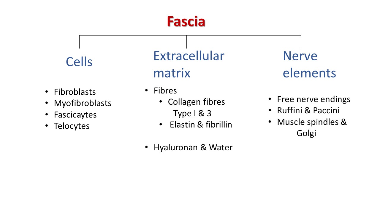
There are different components of the fascia. Each element has a specific role:
- Cells that define the metabolic properties.
- Collagen and elastic fibres are important for the mechanical properties of the tissue.
- Water components and the hyaluronan are defining the capacity of the fascia and the cells.
- Nerve elements define the sensitive role of the fascia.
- Receptors that can change the characteristics of the fascia and they can help the fascia to adapt to different conditions in a physiological situation but also in a pathological situation.
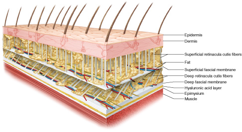
A typical fascia that is composed of two or three layers of parallel collagen fibre bundles separated by thin layers of loose connective tissue. In the dense connective tissue, we can find cells and collagen fibres. In the loose connective tissue, we can also see some vessels, adipocytes, and some other different cells.
The fibrous component of the fascia is involved in force transmission.
The loose connective tissue that is rich in hyaluronan, allows the gliding of the fascia with the muscle but also facilitates the gliding of all sub-layers of the fascial tissue.
Cells in Fascia
There are several specialised cells in fascia:
(a) Fibroblasts
The typical cells of the fascia are the fibroblasts. The fibroblasts are the most important cells of the connective tissue. Fibroblasts maintain the structural integrity and the organization of the connective tissue. They have the role in synthesizing the fibrous components, collagen, and elastic fibers, that play an essential role in regulating force transmission.
(b) Fasciacystes
New findings from the Stecco lab found that there are some cells in the fascia that are different from fibroblasts. Using transmission electron microscope, it is confirmed that these cells are different from the fibroblasts. The fibroblasts are typical elongated cells with collagen fibres. The cells called fasciacytes is different morphologically, and has a different function. The nucleus is round and is very big and clear in the cell. The matrix around these cells is also different. These cells are abundant in hyaluronan. They are usually and the borders of the fascial sub-layers because they can synthesise hyaluronan and so to permit the gliding of the sub-layers of the fascia.
These fasciacytes are fibroblasts-like cells, but they are in a different stage of their life. They become similar to the chondrocytes, but they aren’t chondrocyte because they don’t express the typical marker, but they have the morphology. In a normal healthy fascia, we found about 30% of the cells are fasciacytes, but this percentage can change according to different conditions and in pathological situations.
The fasciacytes are cells that are devoted to the production of hyaluronan. Since hyaluronan is essential for fascial gliding, regulation of these cells could affect the functions of fascia so they could be involved in myofascial pain.
If the fascia is stimulated in a specific way, these cells can increase or decrease their number. For example, if we perform some loading exercise, strength exercise, we can stimulate more the fibroblast, and so the fibroblast can produce and reinforce the tissue producing more collagen fibers. But if we can perform other kinds of movement like a twist, we can stimulate more of the fasciaccytes. We can help the tissue to synthesize more hyaluronan and to permit a correct gliding of the fascia sub-layers.
(c) Myofibroblasts
In the fascia, there are also myofibroblasts that are specialized connective tissue cells with some contractile properties. They are between fibroblasts and muscle cells, and they have this contractual behaviour. They can have a role in the biomechanical functioning of the tissue, and they can regulate the fascia stiffness.
(d) Telocytes
Telocytes are another type of specialized connective tissue. They were discovered for the first time in 2015 by a group in Poland. They define these to the sides like a network in the network system. They have long thin extensions called telopodes. Their specific role is still under investigation, but authors think that they allow intercellular communication. They are found in the tensor fasciae latae, crural fascia of the leg, plantar fascia, and also in the thoracolumbar fascia.
Nervous fibres
For example, in the thoracolumbar fascia of a mouse, nervous fibres follow the fascia like a dense network, thus have a role in pain generation.
Each cell has a specific role in the fascia, but there are still other cells that haven’t been discovered.
In a summary
- Fibroblasts: synthesize the fibrous component regulating the force transmission
- Fasciacytes: synthesize hyaluronan, permitting the gliding of the fascia
- Myofibroblasts: have a contractile property and a role in musculoskeletal dynamics and regulation of fascial stiffness
- Telocytres: have a role in intercellular communication maybe also in the cell repair
- Nervous fibres: involve in the conduction of signals, and have roles in proprioception and nociception
The extracellular matrix
Around all these cells, there is also the extracellular matrix. It divided into a fibrous component and a water component. The fibrous component is made of
- collagen type I: mechanical properties of tensile strength
- collagen type III: reticular fibres arranged in a mesh
- elastic fibres, made up of elastin and fibrillin: have a role of counterbalancing the amount of collagen.
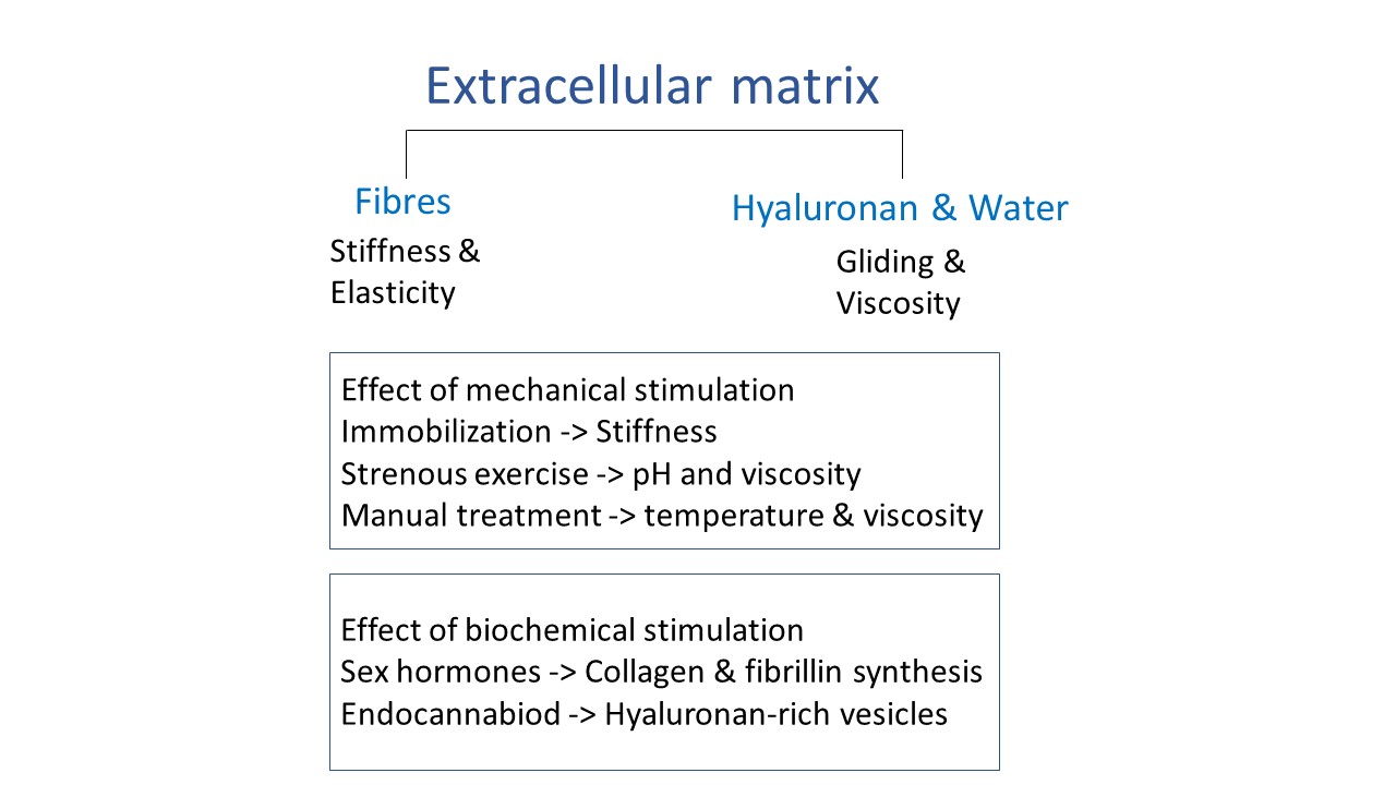
They have a role in transmitting the muscular force. The different amount of collagen and elastic fibres can change according to the anatomic site, the type of fascial tissue, but also different stimuli (hormones, chemical, or mechanical stimuli).
The water component of the extracellular matrix is made of an active network of amorphous materials composed of glycosaminoglycans and proteoglycans. We know that the most critical aminoglycosides, the hyaluronan. Hyaluronan is a very simple molecule but has complex roles.
Hyaluronan (HA) can be found between deep fascia and muscle. HA facilitates gliding between these two structures, and also within the loose connective tissue of the fascia, guaranteeing the smooth sliding of adjacent fibrous fascial layers. It also promotes the functions of the deep fascia. HA can also be found in the endomysium that surrounds the muscles fibres. HA has a rapid turnover in our bodies.
HA can have different roles and opposite roles according to the molecular weight. In general, when HA is in a high molecular weight, they have positive roles: reduce the apoptosis and can decrease the tumour growth and decrease inflammation. But when HA is at a low molecular weight form, it becomes pro-metastatic pro-inflammatory. So, it is not so easy to understand and to study these molecules. We can think hyaluronan is like a sponge and its relation to water. We have two aspects: the molecular weight and concentration. When the molecular weight is big, it is better. But for the concentration, if the concentration is too high, such as in some pathological conditions like immobility or inflammation, the viscosity of the hyaluronan can increase. It means that the HA starts to have a lot of cross-links and a lot of binding of proteins. The water link to the HA molecules decreases and so the tissue starts to be more rigid. The gliding between tissue decreases, and there are much more stiffness and rigidity of the tissue. But in general, in a healthy fascia tissue, the content of HA is always the same.
The mean content of the HA in the fascia is not so high in the fascia; it is the minimum content that permits the gliding. For example, in the skin is much higher than the fascia. But the interesting point is that hyaluronan is always constant in the same kind of fascia. It changes according to the anatomical site.